 |
|
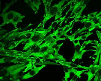
| Fig.2: Detection of VSV mRNA in infected BHK-21 cells using PNA |
Mechanisms of Viral Entry and Replication |
Viral infections are spread all over the world and cause a variety of
diseases. Understanding the molecular mechanisms of viral replication inside the
host cell provides the basis for the development of new therapeutic treatments.
This Project is focused on the genesis of enveloped viruses. An early
consequence of virus infection is the formation of viral mRNA. To follow both
its formation and intracellular location we utilize PNA (Peptide Nucleic Acid)
molecules, tagged with a fluorescent moiety. Upon complementation of the PNA
with the respective mRNA sequence, intercalation of the fluorophore is
accompanied by strong enhancement of quantum efficiency, which can be detected
by (Confocal Laser Scanning) Fluorescence Microscopy in living infected cells.
The advantage of these RNA probes is the stability of the peptide backbone. We
studied the infection of BHK-21 cells by Vesicular Stomatitis Virus (VSV) as a
model system for this technique (Fig. 2). VSV is a negative-sense
single-stranded RNA virus that causes a zootic vesicular disease. This virus
offers several advantages: (i) the existence of highly conserved sequences in
the N- and L- gene as targets for PNAs, (ii) a cytosolic mechanism of
transcription and replication and (iii) the convenience of simple cultivation
and production of infective particles with high titres in different cell lines.
Employing time-lapse imaging we could follow the production of VSV mRNA in
infected cells.
|
|
|
| Fig.2: Detection of VSV mRNA in infected BHK-21 cells using PNA |
References: